 Download files
 Verified to work with the
  To install the software needed to reproduce this system with the
 To set up the environment on the UCSF Wynton cluster to run
this system, run:
To set up the environment on the UCSF Wynton cluster to run
this system, run:
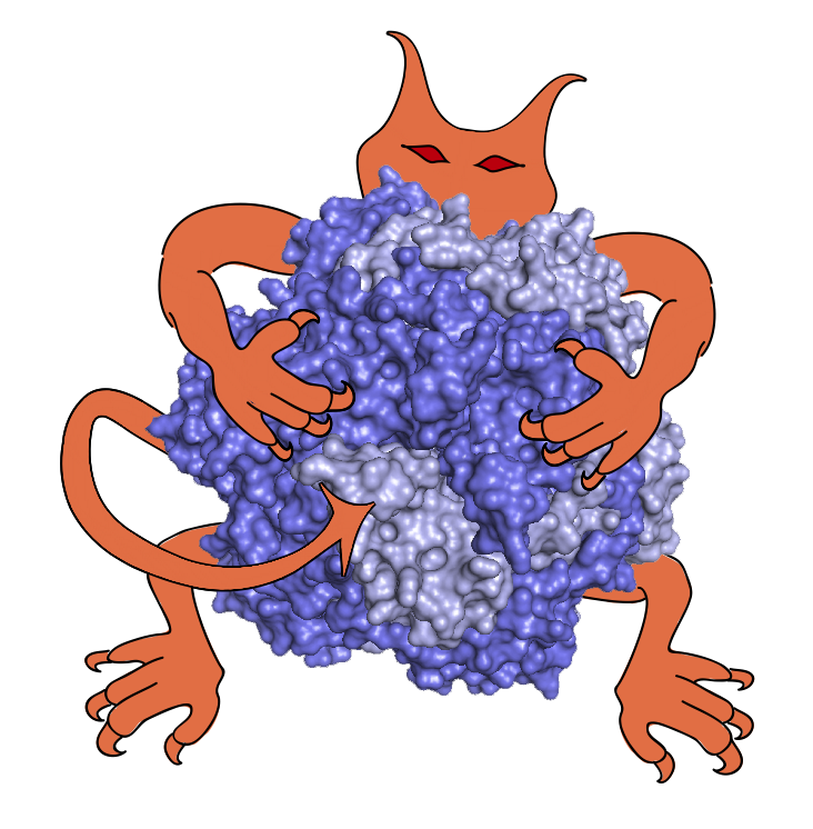
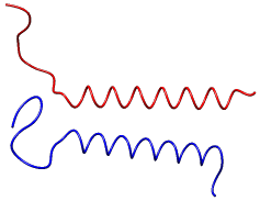
This demonstrates the use of IMP and PMI in the modeling of human glycophorin. The glycophorin is modeled partly with low-resolution beads, and partly as idealized helices. We attempt to reconstruct the native structure (PDB entry 1afo) using cysteine cross-link data [1].
To run, simply run the glycophorin_modeling.py Python script.
On successful completion, output files (the top-scoring models in PDB format,
a trajectory in RMF format, and
statistics files) will be found in the output directory. See the
IMP tutorial
for a full description of these outputs.
[1] Zhu J, Luo BH, Barth P, Schonbrun J, Baker D, Springer TA. Mol Cell. 2009 Apr 24;34(2):234-49. doi: 10.1016/j.molcel.2009.02.022
Author(s): Riccardo Pellarin
License: LGPL. This library is free software; you can redistribute it and/or modify it under the terms of the GNU Lesser General Public License as published by the Free Software Foundation; either version 2 of the License, or (at your option) any later version.
Last known good IMP version: